Specialist in Gynecology and general and obstetric ultrasound in Caballito
ULTRASOUNDS:
- Mammary
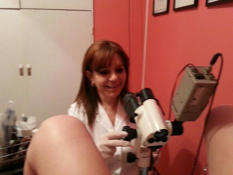
- Gynecological
- Transvaginal
- Abdominals
- Renal
- Liverworts
- Vesiculares
- Pancreáticas
- Hepatobiliopancreática
- Ultrasound Obstetric Controls:
* First trimester
Between week 1 to 10:
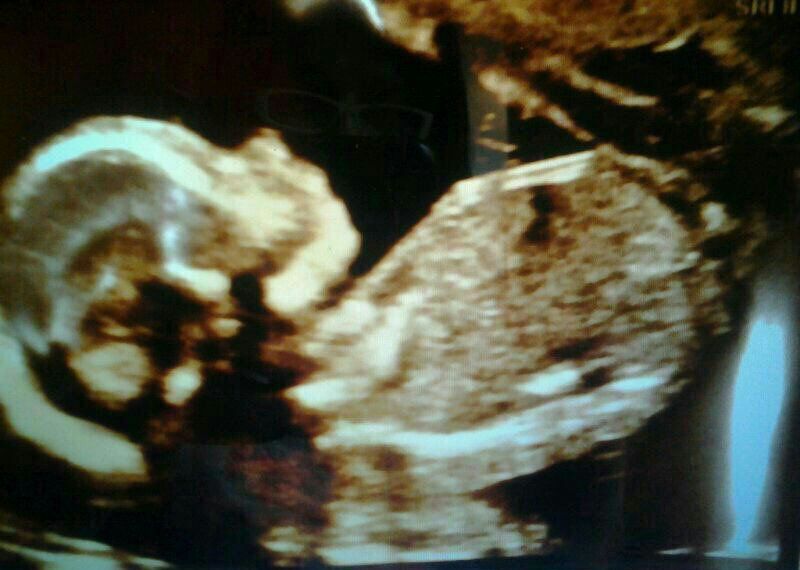
The implantation and vitality of the embryo are monitored.
Gestational age is estimated very accurately according to LMP
(date of last menstruation), which will allow us to evaluate the correct development and growth of the fetus throughout the course of the pregnancy.
We will be able to document whether it is a single or multiple pregnancy (in this case whether it is bi or mono amniotic)
Week 11 to 2:
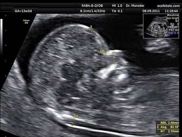
NUCHAL TRANSLUCENCE _(TN)
Through this study (measurement of the transnuchal fold) we attempt to evaluate very precise anatomical structures that suggest the presence of chromosomal abnormalities, for example Down syndrome.
In addition to the TN, we complement this study with other useful ultrasound signs for this purpose such as the evaluation of the nasal bone (NH), Ductus Venosus, Maxillofacial Angle and Tricuspid Regurgitation.
* Second Quarter
Between week 20 to 24:
SCAN FETAL
- At this time, a detailed anatomical study of all the structures of the head, neck, thorax (heart and lungs), abdomen, pelvis and both fetal extremities (humerus, radius, ulna, hand, femur, tibia, fibula and foot) is performed.
*Third Quarter
It takes place between week 25 and week 40:
ECODOPPLER FETAL
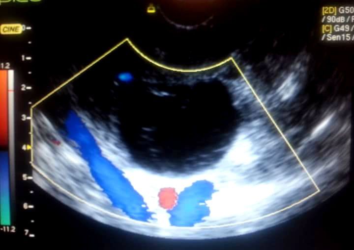
Fetal vitality, assessment of the umbilical cord flow velocity waveform (presence of three vessels, namely two arteries and one vein), the placenta (maturity) and assessment of the uterine arteries (timely decrease in the Resistance Index from the wave of trophoblastic invasion)
Assessment of the quantity of amniotic fluid (Amniotic Fluid Index ILA) and its quality.
It is used to measure and evaluate the blood flow that the baby receives through the umbilical cord, as well as its correct distribution within the baby's body.
It is very useful when fetal growth retardation (IUGR) is suspected, as well as in pregnancies complicated by maternal hypertension, diabetes, thrombophilias and more complex diagnoses, for example, in monoamniotic twin pregnancies, the diagnosis of transfuser-transfused fetus syndrome).
Timely diagnosis of fetal distress.
Between weeks 32 and 36:
AMNIOTIC FLUID MEASUREMENT
Amniotic fluid will reach its maximum volume (800 ml to 1 liter). After this moment, the fluid begins to gradually decrease until it reaches approximately 600 ml at term (40 weeks of gestation).
Both excess (polyhydramnios) and decrease (oligohydramnios) suggest abnormalities that are useful to document using different indices measurable by ultrasound (ILA).
Address: Rivadavia 4704, 4th floor, apt. G
Caballito CITY OF BUENOS AIRES 1424
Email:
informes.cgg@gmail.com
WhatsApp: 54911 61330193


